BARYLITE
Be2Ba(Si2O7),
barium beryllium silicate
Barylite was found in 1929 on the picking table of the Franklin mine, the second locality for the mineral after Långban, Sweden. As Palache (1935) states, barylite from Franklin first attracted attention because of its “vivid blue fluorescence” under the iron arc, and was later found “in pillar 960, 20 feet below the 400-foot level north and about 15 feet from the hanging wall.” Franklin barylite was first studied by Lawson Bauer, who recovered small rectangular plates and fragments of barylite (BYL3) for analysis by dissolving the calcite matrix of what he called the type specimen from Franklin (BYL2). In 1930 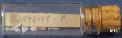 Palache and Bauer described barylite and beryllium-rich vesuvianite in The American Mineralogist under the title, “On the occurrence of beryllium in the zinc deposits of Franklin, N.J.”
Five years later, Palache’s monograph, The Minerals of Franklin and Sterling Hill, New Jersey, settled the description of barylite in the minds of collectors with easily observable features: the mineral was white with prominent cleavage, very hard (Mohs 7), and exhibited “vivid blue fluorescence” under ultraviolet light. The rest of Palache’s description is also worth repeating: “The specimens seen by the author are clearly part of a well-layered vein, the succession of layers from a surface of ore being brown calcite and native copper; gray calcite; a thin zone of willemite and serpentine; white calcite in curved rhombohedrons, strongly fluorescent in ultraviolet light; and barylite, hedyphane, and willemite.”
The best specimens of Franklin barylite probably came from 960 pillar, though this is not known with certainty; in any case there cannot have been many of them. These have barylite in plates 1 cm wide and as much as 10 cm long that diverge like the ribs of a partly opened fan (BYL5). The barylite matrix, as mentioned, consists of calcite, serpentine, hedyphane, copper, and willemite. Large specimens of good quality were rare at the outset and apparently went into university and museum collections such as those at Harvard and Princeton; few are in private hands. Less conspicuous barylite specimens, like those pictured here (BYL1, BYL2) were in private collections but often overlooked; by about 1960 most of the legitimate barylite in Franklin was in several small glass bottles (BYL3), the fragments and plates etched out of matrix 30 years earlier by Lawson Bauer. This does not mean specimens labeled barylite were rare, since any mineral with Parker Shaft credentials that fluoresced vivid blue and was harder than a knife-blade was assumed to be barylite. Examples of faux barylite were in the Kraissl collection, the Areson collection, and many others. That those specimens did not match Palache’s description of barylite’s matrix was widely disregarded, and erroneous descriptions of barylite appeared in amateur and professional mineral publications of the period. Almost all such specimens are now known to be blue-fluorescing margarosanite, disseminated in white barium-rich feldspar which a knife-blade is unlikely to scratch.
The discrepancy between supposed barylite and authentic barylite was apparent to a minority of collectors, but not resolved in print until “The Fluorescence of Barylite from Franklin, New Jersey,” by Richard Gaines and Richard Bostwick, appeared in the Autumn/Winter 1993 issue of The Picking Table. To summarize, the “vivid blue fluorescence” reported by Palache and Bauer in 1930 was seen under the unfiltered iron-arc units which were in wide use at the time; only later, beginning in the mid-1930s, were they gradually replaced by the filtered mercury-arc ultraviolet lamps still in use today. An unfiltered iron arc, not unlike the welder’s arc our parents told us not to stare at, emits violet and blue visible light, plus ultraviolet light below the 254 nm threshold of mercury-arc lamps; in fact, the emission lines of iron at 238-240 nm cause brighter fluorescence in barylite than the 254 nm emission line of mercury. The actual fluorescence of barylite peaks in the longwave ultraviolet region, and the visible portion of its emission spectrum is violet, similar to the fluorescence of hardystonite. However, the perceived fluorescence of barylite under an unfiltered iron arc is a mixture of the violet fluorescence emitted by the mineral, and large amounts of blue and violet light emitted by the iron arc and reflected from the barylite. The fluorescence of Franklin barylite under a filtered mercury-arc shortwave ultraviolet lamp is weak violet, easy to miss in the presence of willemite and calcite. Authentic barylite is fairly easy to identify in hand specimens, once you have read Palache’s full description and, better yet, examined the real thing. If you have access to an iron-arc unit, so much the better. Another diagnostic feature of true barylite is that many specimens include yellow hedyphane, which fluoresces weak orange under midwave ultraviolet light.
References
Abrashev, K.K. and Belov, N.V. (1964), Crystal structure of barylite, BaBe2Si2O7. Doklady Akademii Nauk SSSR, vol. 144, no. 3, p. 636-638.
Abrashev K.K., Ilyukhin V.V., and Belov N.V. (1965), Crystal structure of barylite, BaBe2Si2O7. Soviet Physics - Crystallography, vol. 9, p. 691-699.
Bostwick, R.C. (1977), The fluorescent minerals of Franklin and Sterling Hill, New Jersey: A progress report for 1977, part II. The Picking Table, vol. 18, no. 2, p. 11-24. [Mentions that many specimens labeled "barylite" in area collections are mislabeled and are margarosanite disseminated in feldspar instead; see p. 15-16]
Bostwick, R.C. (1992), The check list of Franklin-Sterling Hill fluorescent minerals: discussion and update. The Picking Table, vol. 33, no. 2, p. 4-11. [Two-line description of barylite fluorescence on p. 11]
Bostwick, R.C. (2016), Fluorescent Minerals of Franklin and Sterling Hill, N.J., Part 1. The Picking Table, vol. 57, no. 2, p. 10-22. [Describes and compares fluorescence of barylite under the iron arc to that under modern shortwave ultraviolet lights on p. 13]
Cannillo, E., Dal Negro, A., and Rossi, G. (1970), On the crystal structure of barylite. Rendiconti della Societa Italiana di Mineralogia e Petrologia, vol. 26, no. 1, p. 53-62.
Dunn, P.J. (1995), Franklin and Sterling Hill, New Jersey: the world’s most magnificent mineral deposits: Privately published in five volumes, 755 pp. [Good description of barylite on p. 407]
Edwards, F.Z. (1960), Barylite. The Picking Table, vol. 1, no. 2, p. 11. [Two-sentence mention of lavender fluorescence of barylite under the iron arc but absence of fluorescence under the shortwave and longwave ultraviolet lights then in use]
Edwards, F.Z. (1963), Barylite - BaBe2Si207 - Hey #14.3.8. The Picking Table, vol. 4, no. 1, p. 5-6. [Reprints passages from the Heinrich and Deane (1962) paper on barylite from Labrador and speculates that lead might be the activator of fluorescence in Franklin barylite based on a published study of synthetic barium silicate phosphors]
Edwards, F.Z. (1972), The fluorescent minerals of the Franklin/Ogdensburg area. The Picking Table, vol. 13, no. 2, p. 11-13. [Notes on p. 11 that Franklin barylite fluoresces “pale to vivid blue” SW. Probably refers instead to margarosanite disseminated in feldspar, a common mistake of the time. See Gaines and Bostwick, 1993]
Gaines, R.V. and Bostwick, R.C. (1993), The fluorescence of barylite from Franklin, New Jersey. The Picking Table, vol. 34, no. 2, p. 22-31. [The definitive paper on local barylite. Provides chemical analyses of four specimens of Franklin barylite, shows fluorescence emission and excitation spectra, describes barylite’s physical properties and associated minerals, and discusses the then-common misidentification of margarosanite disseminated in feldspar as barylite]
Ito, J. and Frondel, C. (1966), Syntheses of barylite, strontium-barylite and lead-barylite. Arkiv för Mineralogi och Geologi Stockholm, vol. 4, p. 391-394. [Notes that natural barylite from Franklin fluoresces bright blue, but none of the synthetic barylites showed any visible response under ultraviolet light]
King, V.T., King, N.E., Betancourt, P.P., Bostwick, R.C., Chin, Peter, Hecht, T.J., Kuitems, S.M., Moritz, Harold, Nemetz, J.D., Nikischer, A.J., Sanford, Stephen, Van Fleet, J.A., and Verbeek, E.R. (2021), The Mineralogy of Franklin and Ogdensburg, New Jersey — A Photographic Celebration. Privately published, 1400 pp. [Barylite discussed on p. 14; photographs on p. 57-58, 236, 297-298, 381, and 437]
Palache, Charles (1935), The minerals of Franklin and Sterling Hill, Sussex County, New Jersey. U.S. Geological Survey Professional Paper 180, 135 pp. [Chemical analysis and description of barylite on p. 92. Notes that barylite “has a vivid blue fluorescence in ultraviolet light,” but this description refers to its appearance under the iron arc, not under modern mercury-vapor SW ultraviolet lights]
Palache, C. and Bauer, L.H. (1930), On the occurrence of beryllium in the zinc deposits of Franklin, New Jersey. American Mineralogist, vol. 15, no. 1, p. 30-33. [Announces discovery of barylite at Franklin in August 1929; provides optical and chemical data, plus a physical description of the species]
Robinson, P.D. and Fang, J.H. (1977), Barylite, BaBe2Si207: its space group and crystal structure. American Mineralogist, vol. 62, 167-169.
Images
