DIOPSIDE


CaMgSi2O6,
calcium magnesium silicate


Diopside must surely have been found in some of the earliest openings in the Franklin Marble and appears in early lists of the local minerals, but Palache (1935, p. 62) remarked that all specimens so labeled from old collections had turned out to be some other mineral, and that the first definitive evidence of its presence was an unpublished chemical analysis made in 1926 by Lawson Bauer, chemist for the New Jersey Zinc Company. Despite such early confusion, diopside is a common mineral locally, with numerous authenticated specimens now known.

Diopside from the Franklin Marble is generally close to end-member CaMgSi2O6, is white to nearly colorless, occurs generally in a matrix of coarse-grained marble, and fluoresces bright pale blue under shortwave (SW) ultraviolet (UV) light. The activator of the blue fluorescence is probably titanium (as TiO6 groups), present in the diopside as an impurity. Norbergite and tremolite are the most commonly associated silicate minerals, but numerous other silicates have been found with diopside as well. Most diopside in the Franklin Marble occurs as fairly small (2-5 mm), equant to slightly elongated grains and rough crystals, often scattered within the host marble (DPS11), but in some specimens forming dense aggregates or layers of nearly pure material (DPS20). Specimens of such diopside are familiar to most collectors of the local minerals and have been recovered from numerous exposures of the Franklin Marble throughout its extent, from the Sparta, N.J. area in the south to the Amity, N.Y. area in the north. Similar specimens have been recovered underground in the Franklin and Sterling zinc mines, but only in the Franklin Marble, in areas external to the Zn-Mn-Fe orebodies.

Diopside from the zinc-rich orebodies at Franklin and Sterling Hill is of wholly different character from that in the Franklin Marble. Far from being pure, it often contains appreciable zinc, manganese, and iron, and occurs as tan, brown, or ivory-colored grains and granular aggregates with a pronounced platy appearance (DPS4, DPS7, DPS12) due to closely spaced basal parting surfaces (Palache, 1935, p. 62). Cleavage is also present but in most specimens is visually subordinate to the partings. Grain size varies considerably but is often coarse, with individual grains 2-10 cm across, and some larger. As noted by Dunn (1995, p. 433), commonly associated minerals include andradite, franklinite, willemite, and calcite, and also hardystonite, bustamite, rhodonite, and apatite. In some specimens the parting surfaces bear thin films of nearly colorless to white clinohedrite. Until recently it was thought that diopside from the Franklin and Sterling Hill orebodies does not fluoresce. In 1917-1918, however, several specimens of “white schefferite” (low-iron diopside) from Franklin were observed to fluoresce red under longwave (LW) UV light (Verbeek et al., 1918). The fluorescence is of only dim to moderate brightness, similar in color to that of bustamite, but is unmistakable (DPS17).

The wide compositional range of pyroxenes in the Franklin and Sterling Hill orebodies gave rise to several varietal names that remained in use until relations among the various pyroxene species became better defined. Zinc schefferite and schefferite are the most common of these and were described under those headings by Palache (1935, p. 62-63). Zinc schefferite had earlier been described by Hillebrand (1900) and Wollf and Melczer (1900), and schefferite a decade later by Palache (1910). Numerous specimens in old collections bear such names, and most of them, particularly those of pale to medium brown color, conform to what we now know as diopside. White schefferite is another such term and refers to diopside of overall ivory-white color. A comprehensive revision of the nomenclature of the pyroxenes (Morimoto et al., 1978) led to abandonment of those names, plus more than 100 others, and to the formal acceptance of the species names in use today.

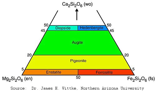
The number of possible elemental substitutions in the pyroxenes, Pyroxene Quadrilateral and the existence of at least partial solid-solution series among many of them, frustrate attempts to identify these minerals to the species level without a comprehensive chemical analysis. With increasing iron content, for example, diopside grades into hedenbergite, and with increasing Mg content it grades into augite. All three species are known from both the Franklin and Sterling Hill deposits. The pyroxene quadrilateral shown to the right depicts the compositional ranges of diopside, augite, and hedenbergite with respect to the pure Ca, Mg, and Fe end members. It is important to realize, however, that additional substituents, most notably Mn and Zn, appear in Franklin-Sterling Hill pyroxenes as well. Dunn (1995) suggested that only light-colored pyroxenes from the Franklin-Sterling Hill area be termed diopside unless proven otherwise. Darker brown to dark green pyroxenes are commonly augite but could also be the sodic (Na-rich) pyroxenes aegirine-augite or aegirine, also known locally. Hedenbergite, due to its high iron content, is dark green to nearly black, but so too is much diopside containing lesser amounts of iron.
References

Betancourt, P.B. (1989), The epidote-pyroxene-fluorapophyllite assemblage in the Franklin mine at Franklin, New Jersey. The Picking Table, vol. 30, no. 1, p. 8-10. [Mentions a pale green pyroxene, regarded as probable diopside, from the title assemblage at Franklin]

Bostwick, R.C. (1977), The fluorescent minerals of Franklin and Sterling Hill, NJ. The Picking Table, vol. 18, no. 1, p. 15-24. [Short description of daylight appearance and fluorescence of diopside from the Franklin Marble on p. 24]

Bostwick, R.C. (1992), The check list of Franklin-Sterling Hill fluorescent minerals:  discussion and update. The Picking Table, vol. 33, no. 2, p. 4-11. [Fluorescence of diopside described in brief on p. 10]

Bostwick, R.C. (2016), Fluorescent minerals of Franklin and Sterling Hill, N.J., Part 1. The Picking Table, vol. 57, no. 2, p. 10-22. [Diopside discussed on p. 20]

Dunn, P.J. (1995), Franklin and Sterling Hill, New Jersey:  the world’s most magnificent mineral deposits. Privately published, 755 p. [Full description of diopside on p. 432-434; chemical analyses of several specimens in Table 10 on p. 430]

Edwards, F.Z. (1972), The fluorescent minerals of the Franklin/Ogdensburg area:  The Picking Table, vol. 13, no. 2, p. 11-13. [Mentions on p. 12 a “good blue-white” fluorescence of local diopside, but this is true only for diopside from the Franklin Marble]

Frondel, Clifford (1972), The minerals of Franklin and Sterling Hill - a checklist. Wiley Interscience, New York, 94 pp. [Good one-paragraph description of diopside on p. 53]

Grenier, Gary (2004), The art of fluorescent mineral photography, with special attention to the minerals of Franklin and Sterling Hill, Part II. The Picking Table, vol. 45, no. 1, p. 9-21. [Brief discussion of fluorescence of diopside from a photographer’s viewpoint on p. 10-11]

Hillebrand, F.W. (1900), Mineralogical notes:  Jeffersonite. U.S. Geological Survey Bulletin 167, p. 68-69.

Johnson, C.A. (1990), Petrologic and stable isotopic studies of the metamorphosed zinc-iron-manganese deposit at Sterling Hill, New Jersey. Ph.D. Dissertation, Yale University, New Haven, Connecticut, 108 pp. [Includes chemical analyses of diopsides from Sterling Hill]

King, V.T., King, N.E., Betancourt, P.P., Bostwick, R.C., Chin, Peter, Hecht, T.J., Kuitems, S.M., Moritz, Harold, Nemetz, J.D., Nikischer, A.J., Sanford, Stephen, Van Fleet, J.A., and Verbeek, E.R. (2021), The Mineralogy of Franklin and Ogdensburg, New Jersey—A Photographic Celebration. Privately published, 1400 pp. [Fluorescence described on p. 17; photos of fluorescent specimens on p. 94-96; others on p. 852-853]

Morimoto, N., Fabries, J., Ferguson, A.K., Ginzburg, I.V., Ross, M., Seifert, F.A., Zussman, J., Aoki, J., and Gottardi, G. (1988), Nomenclature of pyroxenes. The American Mineralogist, vol. 73, p. 1123-1133. [Comprehensive revision of pyroxene nomenclature; led to acceptance of 20 species names and abandonment of 105 superfluous names]

Moritz, Harold and Craig, F.M. (2023), Analyses of clinopyroxenes from Franklin and Ogdensburg, New Jersey, U.S.A. The Picking Table, vol. 64, no. 1, p. 12.

Palache, Charles (1910), Contributions to the mineralogy of Franklin Furnace, N. J. American Journal of Science, 4th Series, vol. 29, p. 177-187.

Palache, Charles (1935), The minerals of Franklin and Sterling Hill, Sussex County, New Jersey. U.S. Geological Survey Professional Paper 180, 135 pp. [Description of diopside and its varieties (schefferite, zinc schefferite) on p. 61-63.]

Reilly, L.C., (1983), An analysis of calc-silicate bands of the Sterling Hill zinc deposit, Sussex County, New Jersey. Senior Thesis, Department of Geology and Geophysics, Yale University, New Haven, Connecticut, 90 pp. [Mentions rhodonite exsolved from diopsidic pyroxene from Sterling Hill]

Verbeek, E.R., Mazel, Charles, and Kuitems, S.M. (2018), Red-fluorescent diopside from the orebody at Franklin, New Jersey. The Picking Table, vol. 60, no. 1, p. 25-27.

Wolff, J.E. and Melczer, G. (1900), On hardystonite and a zinc schefferite from Franklin Furnace, New Jersey, with a note on the optical constants of the schefferite. Proceedings of the American Academy of Arts and Sciences, vol. 36, no. 7, p. 113-118. [Provides a description, chemical analysis, and optical data on newly found specimens of pyroxene from Franklin].


Images

Diopside from the Franklin and Sterling Hill orebodies 

Diopside from the Franklin Marble 


Franklin and Sterling Hill orebodies


Very coarse-grained, medium-brown diopside and white calcite, scattered grains of franklinite, pale tan hardystonite, willemite
Earl R. Verbeek
A large specimen consisting almost wholly of very coarse-grained, medium-brown diopside and white calcite, but including scattered grains of franklinite (most 0.5 cm or less across) and small masses of pale tan hardystonite, plus willemite (fl. green SW) and clinohedrite (fl. orange SW) as minor hydrothermal alteration products. The main diopside grain is 8 cm across and displays prominent basal partings that cause it to break into thin plates. Franklin Mineral Museum specimen FMM-607, ex Ewald Gerstmann collection.

Identifier: DPS4a
Locality: Franklin mine, Franklin
Specimen size: 26 x 18 x 12 cm


Single large grain of brown diopside, nearly white hardystonite, black franklinite, grains of pale green willemite and pink tephroite
Earl R. Verbeek
A portion of a single large grain of brown diopside showing prominent basal partings, which give the mineral a platy structure, here most evident toward the base of the specimen. The diopside is poikiloblastic and encloses numerous grains of nearly white hardystonite and black franklinite, plus (toward right side as photographed) grains of pale green willemite and pink tephroite. This specimen, catalog number FMM-5115, was donated to the Franklin Mineral Museum by Al Standfast, whose skills as a photographer (both of specimens and people) were much appreciated locally.

Identifier: DPS7a
Locality: Franklin mine, Franklin
Specimen size: 17.5 x 10 x 4 cm


Single grain of diopside, scattered grains of black franklinite and pale pinkish-tan grains of hardystonite
Earl R. Verbeek
Part of a single grain of diopside showing well-developed partings parallel to the photographed face. Included in the diopside are scattered grains of black franklinite and pale pinkish-tan grains of hardystonite. The top and bottom surfaces of the specimen are lightly filmed with clinohedrite and a greenish-blue, secondary copper mineral. This handsome example is from the collection of Steven Phillips and previously was specimen no. 571 in the Richard Hauck collection.

Identifier: DPS14a
Locality: Franklin mine, Franklin
Specimen size: 10.5 x 6 x 5.5 cm


Orange-tan, very coarse-grained with granular franklinite ore
Earl R. Verbeek
Orange-tan, very coarse-grained diopside showing pronounced, thinly spaced parting surfaces, in contact with granular franklinite ore at the base. Not visible in this daylight view is abundant secondary willemite as patchy films on the diopside parting surfaces, along with a trace of clinohedrite. Note, in upper right, the numerous franklinite grains included in the diopside. The current whereabouts of this specimen are unknown; when photographed it was part of the sales stock of the Sterling Hill Mining Museum.

Identifier: DPS12b
Locality: Franklin mine, Franklin
Specimen size: 13 x 9 x 8 cm


Coarse-grained, ivory-colored diopside with scattered grains of black franklinite and some brown tephroite
Earl R. Verbeek
Coarse-grained, ivory-colored diopside showing excellent basal parting, with scattered grains of black franklinite and (lower right) some brown tephroite. The visible tephroite is part of a much larger mass exposed mostly on the back of the specimen. Also present is secondary willemite, both as patchy films on parting surfaces and thin veinlets cutting through the mass, but these are apparent only by their fluorescence. Ex John Sanfaçon collection, presently (2025) in the collection of Steven Kuitems. This is the specimen in which red fluorescence in Franklin diopside was first documented, as shown in photo DPS17b.

Identifier: DPS17a
Locality: Franklin mine, Franklin
Specimen size: 9 x 5.5 x 4 cm


Coarse-grained, ivory-colored diopside with scattered grains of black franklinite and some brown tephroite under longwave UV Light
Earl R. Verbeek
This is how the specimen shown in photo DPS17a appears under a longwave ultraviolet LED light source. The red fluorescence almost certainly is due to manganese as the Mn2+ ion substituting for Mg2+ in the diopside structure. The diopside was confirmed as such both by Raman spectroscopy and X-ray fluorescence analysis. The chemical data also confirmed its low iron content (only 0.54 wt.% FeO), consistent with its abnormally pale color. Discussion of this specimen, plus fluorescence emission and excitation spectra of the diopside, were presented in Verbeek et al. (2018). Since publication of that paper, several additional specimens of red-fluorescing diopside from the Franklin orebody have been discovered, all of pale tan to nearly white color.

Identifier: DPS17b
Locality: Franklin mine, Franklin
Specimen size: 9 x 5.5 x 4 cm


Well-formed, brown crystals of diopside 1-3 cm long with franklinite crystals in a matrix of coarse-grained, pale gray calcite
Earl R. Verbeek
Well-formed, brown crystals of diopside 1-3 cm long with franklinite crystals of similar dimension, both in a matrix of coarse-grained, pale gray, brightly red-fluorescing calcite. Originally in the Ewald Gerstmann collection, this is specimen FMM-599 on display in the Franklin Mineral Museum.

Identifier: DPS8a
Locality: Franklin mine, Franklin
Specimen size: 13 x 11 x 5 cm


Pinkish-brown diopside, pale green willemite, dark brown garnet, and subordinate gray tephroite
Earl R. Verbeek
Pinkish-brown diopside (showing well-developed parting) with abundant pale green willemite, dark brown garnet, and subordinate gray tephroite. The parting surfaces in the diopside are liberally coated in places with thin, arborescent films of a black mineral, most likely a secondary manganese mineral or possibly native lead. This attractive specimen is in the Steven Phillips collection. Prior owners include John Blake and, later, Gary Grenier (GG2362), who obtained it in April 1994 from Fred Parker.

Identifier: DPS13a
Locality: Franklin mine, Franklin
Specimen size: 10 x 7.5 x 3.5 cm


Franklin Marble


Thickly scattered grains of very pale mustard-yellow diopside in coarse-grained white calcite marble
Earl R. Verbeek
Thickly scattered grains of very pale mustard-yellow diopside in coarse-grained white calcite marble. The tiny black grains are graphite. Though an obvious color difference exists between the diopside grains and the enclosing calcite, in other specimens the color difference is almost nil and the presence of diopside is readily overlooked. No such problem exists under SW ultraviolet light; see photo DPS11b. This is specimen 3044 of the Mark Boyer collection.

Identifier: DPS11a
Locality: Franklin Marble, Sussex County
Specimen size: 8.5 x 6 x 4 cm


Thickly scattered grains of very pale mustard-yellow diopside in coarse-grained white calcite marble under shortwave UV Light
Earl R. Verbeek
In this view – the same specimen as in photo DPS11a, but under SW ultraviolet light – both the distribution of the diopside grains and their margins against the enclosing marble are distinct. The blue “haze” around some of the diopside grains results from transmittal through the enclosing, slightly translucent calcite of some of the blue light emitted by the diopside.

Identifier: DPS11b
Locality: Franklin Marble, Sussex County
Specimen size: 8.5 x 6 x 4 cm


A thin, platelike specimen of pale gray, coarse-grained, graphitic marble bisected by a layer 2.5 to 3 cm thick of fine-grained, straw-yellow diopside
Amanda Tabares
A thin, platelike specimen of pale gray, coarse-grained, graphitic marble neatly bisected by a layer 2.5 to 3 cm thick of fine-grained, straw-yellow diopside. The diopside fluoresces bright medium blue SW, appearing much like scheelite. This specimen came from the adit level of the Sterling mine during blasting operations for the Edison Tunnel in the 1990s and was acquired at that time by Richard Bostwick, who donated it to the Franklin Mineral Museum on August 4, 2024. It is probably one of those displayed in the large fluorescent tent during the 2007 Springfield, Massachusetts mineral show, when 13 cases of fluorescent minerals were a featured attraction.

Identifier: DPS20a
Locality: Edison Tunnel, Sterling mine, Ogdensburg
Specimen size: 11 x 9.5 x 2.5 cm


A thin, platelike specimen of pale gray, coarse-grained, graphitic marble bisected by a layer 2.5 to 3 cm thick of fine-grained, straw-yellow diopside under shortwave UV Light
Amanda Tabares
Same specimen as in photo DPS20a, showing the bright blue SW fluorescence of the diopside. Though it came from the Sterling mine, this specimen is from an area external to the Sterling Hill Zn-Fe-Mn orebody and thus has the typical appearance and fluorescence of diopside from the Franklin Marble.

Identifier: DPS20b
Locality: Edison Tunnel, Sterling mine, Ogdensburg
Specimen size: 11 x 9.5 x 2.5 cm


Pale yellow grains of norbergite and white to pale gray grains of diopside with small, scattered grains of violet fluorite, graphite, and pyrite
Richard Schnurr (Fox Tail Images)
Pale yellow grains of norbergite and white to pale gray grains of diopside, both common, in marble matrix, with small, scattered grains of violet fluorite, graphite, and pyrite. The diopside is nearly the same color as the matrix rock, so it is inconspicuous in daylight, but its SW blue fluorescence reveals its abundance (see photo DPS18b). The norbergite fluoresces its customary yellow color SW. This specimen was purchased during the early 2000s by Richard Bostwick from Lenny Lee at one of the local mineral shows and later, in 2007, was exhibited in the large fluorescent tent at the Springfield, Massachusetts show. In 2023 it became specimen FMM-3150 of the Franklin Mineral Museum through purchase of a large part of the Bostwick collection.

Identifier: DPS18a
Locality: Franklin Marble, Sussex County
Specimen size: 13 x 10 x 6 cm


Pale yellow grains of norbergite and white to pale gray grains of diopside with small, scattered grains of violet fluorite, graphite, and pyrite under shortwave UV Light
Richard Schnurr (Fox Tail Images)
Viewed under SW ultraviolet light, this combination of blue-fluorescing diopside and yellow-fluorescing norbergite (same specimen as in photo DPS18a) is unusually attractive. Such "half-and-half" specimens, rich in both minerals and nicely separated into blue- and yellow-fluorescing portions, are uncommon.

Identifier: DPS18b
Locality: Franklin Marble, Sussex County
Specimen size: 13 x 10 x 6 cm


Two halves of a stream-rounded cobble consisting mostly of diopside with a little calcite
Earl R. Verbeek
Two halves of a stream-rounded cobble consisting mostly of diopside with a little calcite. The adhering bits of sand show it was collected not from a modern stream bed, but from one of the sand-and-gravel pits that exploit the local glacial outwash deposits, where rainwater descending through the permeable sediments gradually deposits films of calcite that cement some of the grains together. This is an uncatalogued specimen of the Franklin Mineral Museum.

Identifier: DPS9a
Locality: A sand and gravel pit in the Franklin-Ogdensburg area of Sussex County.
Specimen size: 12 x 9 x 8 cm


Two halves of a stream-rounded cobble consisting mostly of diopside with a little calcite
Earl R. Verbeek
The two halves of the glacial cobble shown in photo DPS9a, separated to show the interior. Most of the cobble consists of massive, faintly yellow diopside, bordered on one side (upper left as shown) by coarse-grained, pale gray calcite containing scattered, tiny flecks of graphite.

Identifier: DPS9b
Locality: A sand and gravel pit in the Franklin-Ogdensburg area of Sussex County.
Specimen size: 12 x 9 x 8 cm


Two halves of a stream-rounded cobble consisting mostly of diopside with a little calcite under shortwave UV Light
Earl R. Verbeek
The granularity and richness in diopside of the glacial cobble shown above (photos DPS9a and DPS9b) is most evident under SW ultraviolet light, as seen here. The thin, nonfluorescent border on the upper left of each half of the cobble is calcite marble.

Identifier: DPS9c
Locality: A sand and gravel pit in the Franklin-Ogdensburg area of Sussex County.
Specimen size: 12 x 9 x 8 cm


Myriad small crystals of pale gray diopside, pale brown phlogopite, and subordinate violet fluorite
Earl R. Verbeek
Myriad small crystals of pale gray diopside, pale brown phlogopite, and subordinate violet fluorite, the whole dissolved from the host calcite. The Franklin Marble in places was so silica-rich that calcite was a subordinate component of the rock and could be dissolved in acid to produce a skeletal framework of other minerals, as exemplified here. This peculiar specimen was obtained by Gary Grenier in July 1990 from Al Bliss and entered the Grenier collection as specimen GG2524. It later became part of the Steven Phillips collection.

Identifier: DPS16a
Locality: Franklin Marble, Sussex County
Specimen size: 8 x 6.5 x 5 cm


Pale greenish-gray, prismatic diopside crystals, most of them 2-3 cm in length
Earl R. Verbeek
A small group of pale greenish-gray, prismatic diopside crystals, most of them 2-3 cm in length, naturally weathered from marble matrix. An old, discolored paper label with the specimen gives, in pencil, the locality as "Hardingston” [sic], New Jersey, a misspelling of the colonial land tract of Hardiston, established by Royal Charter in 1762. Originally this tract encompassed most of eastern Sussex County, including present-day Hardyston, Vernon, and Sparta Townships, so the exact locality of this specimen is lost to history. This intriguing piece is from the Steven Phillips collection. Though it has not been analyzed, the pale greenish color suggests a low iron content, consistent with the suggested identity of diopside.

Identifier: DPS15a
Locality: Franklin Marble, Sussex County
Specimen size: 6 x 3.5 x 3 cm